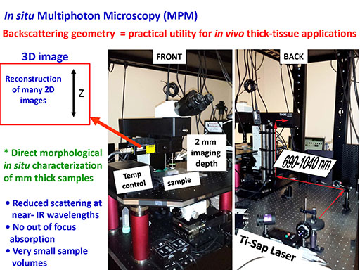

A "work-horse" multiphoton imaging system used regularly and maintained at UC Riverside by our laboratory is an open space upright Thorlabs Multiphoton Laser-Scanning Microscope. It is based on the Nikon microscope body and features a standard illumination system for the transmitted light and a mountable LED ring light for reflected illumination. The femtosecond Ti:Sapphire laser from Spectra-Physics is coupled into the microscope and provides femtosecond pulses at a repetition rate of about 80 MHz, with the center frequency tunable from 690 nm to 1040 nm. Objectives focus the light and allow tightly focused femtosecond pulsed radiation to interact with samples. The backscattered signals thus generated are collected by the same objectives, transmitted through the appropriate dichroic mirrors and barrier filters, separated and refocused on the photomultiplier tube (PMT) detectors. Low noise and high quantum efficiency photon counting detectors (Hamamatsu H7422-40 PMT) were selected for this application. The output of the PMTs is converted to photoelectron pulses. The data acquisition process and the scanner movement are synchronized by the software to generate 2D images that are reconstructed into 3D images by the computer.
The Multiphoton Laser-Scanning Microscope features an aluminum physiology stage consisting of a rigid, mechanically and thermally stable breadboard (U-shaped, 650 mm x 450 mm x 25 mm) with 1/4-20 tapped individually sealed mounting holes. The breadboard is supported by four support columns that can be locked into place and attached to the optical table. Long working distance (between 2 mm and 2.6 mm imaging range) water immersion objectives Zeiss, 63x water, N.A. 1.0; Zeiss 10x water, N.A. 0.4 and Olympus 20x water, N.A.1.0 are aberration corrected and suitable for UV-VIS/nearIR imaging/excitation.
The optical setup is easily customizable to fit needs of a specific imaging experiment including laser polarization experiments. In-house build temperature controlled stage adaptor made from anodized aluminum is available to maintain physiological temperature of the biological systems. It is about 250 mm x 200 mm x 1.5 mm with adhesive silicone heating pads underneath. The temperature of the heating pads is controlled through the resistive heating by the Omega Engineering microcontroller with a thermocouple (type J, SA1-J) feedback.
MPM combines the advantage of visualization at a high spatial resolution (transverse: ~0.5 micrometer; axial: ~1.5 micrometer) with high contrast and functional information available from spectroscopy that we perform as well.
MPM uses near infrared light to generate intrinsic two-photon excited fluorescence (TPF) from UV/VIS absorbing molecules and proteins and/or second harmonic signals (SHG) from structures without a center of symmetry such as collagen.
Our choice of the wide tuning range frequency Ti:Sapphire laser (fs, 690 to 1040 nm, bandwidth ~ 10 nm) enables visualization of many induced and intrinsic fluorescent cross-links and their in-situ characterization along with monitoring self-assembly of collagen states within biological materials.
Design: J.G.L
Copyright 2007 J.G.L. All rights reserved.